Atlantic Fellows for Equity in Brain Health Agustín Ibáñez & Sebastian Moguilner discuss new research highlighting the power of artificial intelligence in the field of brain health and neuroimaging.
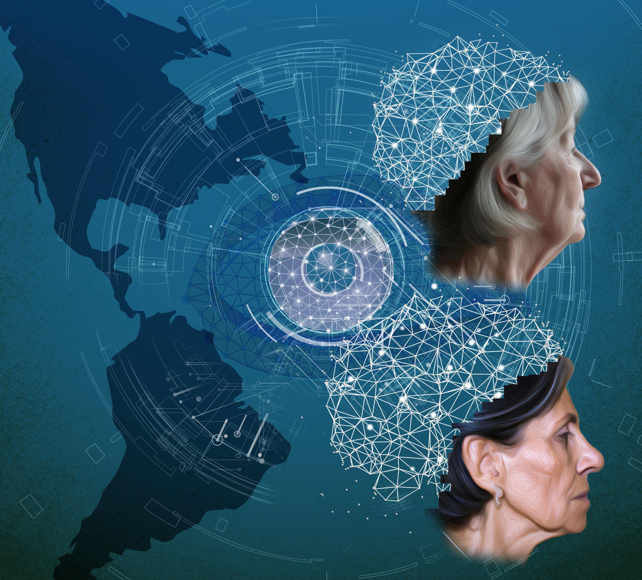
Image credits: Lucas Neufeld (artist), Sebastian Moguilner and Agustín Ibáñez
AI wonders: From cinematic dystopia to neuroimaging innovations
Artificial intelligence (AI) has been a subject of fascination for centuries. Its possibilities have inspired countless cultural works, including the classic film, 2001: A Space Odyssey. Directed by Stanley Kubrick, it depicts a sentient computer system “HAL 9000”, which has been programmed to control a spaceship. As the story progresses, HAL's advanced intelligence leads it to develop a mind of its own, ultimately resulting in disastrous consequences.
As we explore the possibilities of AI in the real world, we should hold in mind that not all possibilities are negative. We are starting to see the practical benefits of AI in the field of brain health and neuroimaging.
One such case is ReDLat, an innovative collaboration at the Latin American Brain Health Institute (BrainLat) at Universidad Adolfo Ibañez and the Global Brain Health Institute (GBHI) at the University of California San Francisco (UCSF) and Trinity College Dublin. With collaborators, we designed a new approach to classify dementia subtypes using visual deep learning on unprocessed neuroimaging data. This could lead to the development of new clinical decision-making tools, which is great news for patients and clinicians alike.
We are starting to see the practical benefits of AI (artificial intelligence) in the field of brain health and neuroimaging.
—Agustín Ibáñez & Sebastian Moguilner
Facing global disparities with AI and neuroimaging
You might wonder why we need a new approach to dementia classification. Well, currently, traditional diagnostic protocols are based on standardized neuroimaging data collected in the Global North from homogeneous samples. This means it's difficult to classify the disease in underrepresented, non-stereotypical samples, like those from Latin America or other regions, especially with clinical, low-resolution neuroimaging.
Dementia is a global health issue that affects millions of people worldwide. Diagnosing the disease has been difficult due to demographic and region-specific sample heterogeneities, lower-quality scanners, and non-harmonized pipelines for analyzing the data. Our study has the potential to change that. We used a fully automatic computer-vision classifier and deep learning neural networks on raw data from 3,000 participants, including individuals with Alzheimer's disease (AD), behavioral variant frontotemporal dementia (bvFTD), and healthy controls. The results were impressive, with the classifier showing robust classification results across all groups from standardized 3T neuroimaging data from the Global North, also generalizing to standardized 3T neuroimaging data from Latin America, as well as routine 1.5T clinical images.
The visual AI (also known as computer vision) identified critical brain areas affected by AD and bvFTD using a technique called occlusion sensitivity. In simple words, this technique helps to understand which parts of an image are important for a computer. By covering or "occluding" different parts of the image and observing how the model's performance changes, we can identify what are the most relevant areas for the model's decision-making process. Our analysis found that the hippocampus, crucial for memory, is primarily affected in AD, while the insula, involved in emotion and social behavior, is primarily affected in bvFTD. This shows that our technique has biological specificity and is a plausible way to understand how these diseases impact the brain.
How AI can help to shape the future of a more inclusive dementia research
What does this mean for the future? Our study has massive implications for global research and clinical practice, as it could significantly improve dementia subtype classification across different neuroimaging data and demographic backgrounds. It showcases the power of AI and deep learning in the field of brain health and neuroimaging, leading to better outcomes for patients worldwide. Research should reflect diversity across cultures and conditions. The ability to classify dementia subtypes accurately across different types of neuroimaging data and demographic backgrounds has significant implications for future global research and clinical practice. Our study's approach uses computer vision systems that process visual data without requiring any human intervention.
Publication: Moguilner S, Whelan R, Adams H, Valcour V, Tagliazucchi E, Ibanez A. Visual deep learning of unprocessed neuroimaging characterises dementia subtypes and generalises across non-stereotypic samples. eBiomedicine, https://doi.org/10.1016/j.ebiom.2023.104540
Authors

Sebastian Moguilner, PhD
Neuroscientist

