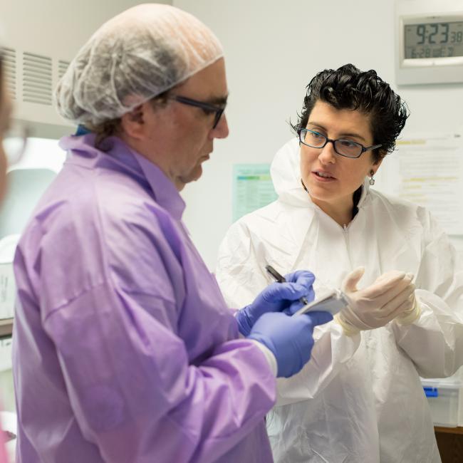
Atlantic Fellow Ignacio Illán-Gala discusses the use of biomarkers to improve the accuracy and scalability of dementia diagnoses.
The diagnosis of neurodegenerative dementias can be challenging because some of the first symptoms can be mild or resemble other psychiatric conditions, like depression or bipolar disorder. In addition, the same symptoms can be associated with different causes of dementia that may benefit from different treatments. However, biomarkers may be key to improving the accuracy and scalability of dementia diagnoses.
What are biomarkers?
Biomarkers are biological molecules found in blood, other body fluids, or tissues that are a sign of a normal or abnormal process, or of a condition or disease. They are powerful tools allowing early diagnosis of neurodegenerative dementias. They can be used for four primary purposes:
- Guide clinical diagnosis by identifying the key functional changes of a disease (so-called diagnostic markers)
- Estimate the risk or speed of progression of a disease (prognostic markers)
- Monitor the progression or response to therapy (theragnostic markers)
- Characterize the specific cause of dementia (i.e., Alzheimer´s disease)
However, some biomarkers may be too expensive for low and middle-income countries, while others are not appropriate for broad use because of lack of diversity considerations (e.g., sex-related differences).
Creating new imaging biomarkers to improve early detection
As an Atlantic Fellow for Equity in Brain Health now based in Barcelona (Spain), I am determined to create new imaging biomarkers to improve the early detection of neurodegenerative dementias. As a neurologist, I work on the diagnosis and treatment of patients with frontotemporal dementia. My research is focused on validating new tools aiming to increase diagnostic precision and improve our ability to understand how the disease progresses. I believe this is an essential step toward the development of effective treatments.
Using a Pilot Award for Global Brain Health Leaders, I investigated the potential value of a new imaging biomarker that is more sensitive than conventional Magnetic Resonance Imaging (MRI) to the earliest changes in neurodegenerative dementias. Read more about my pilot project, Microstructural Changes in Primary Progressive Aphasia.
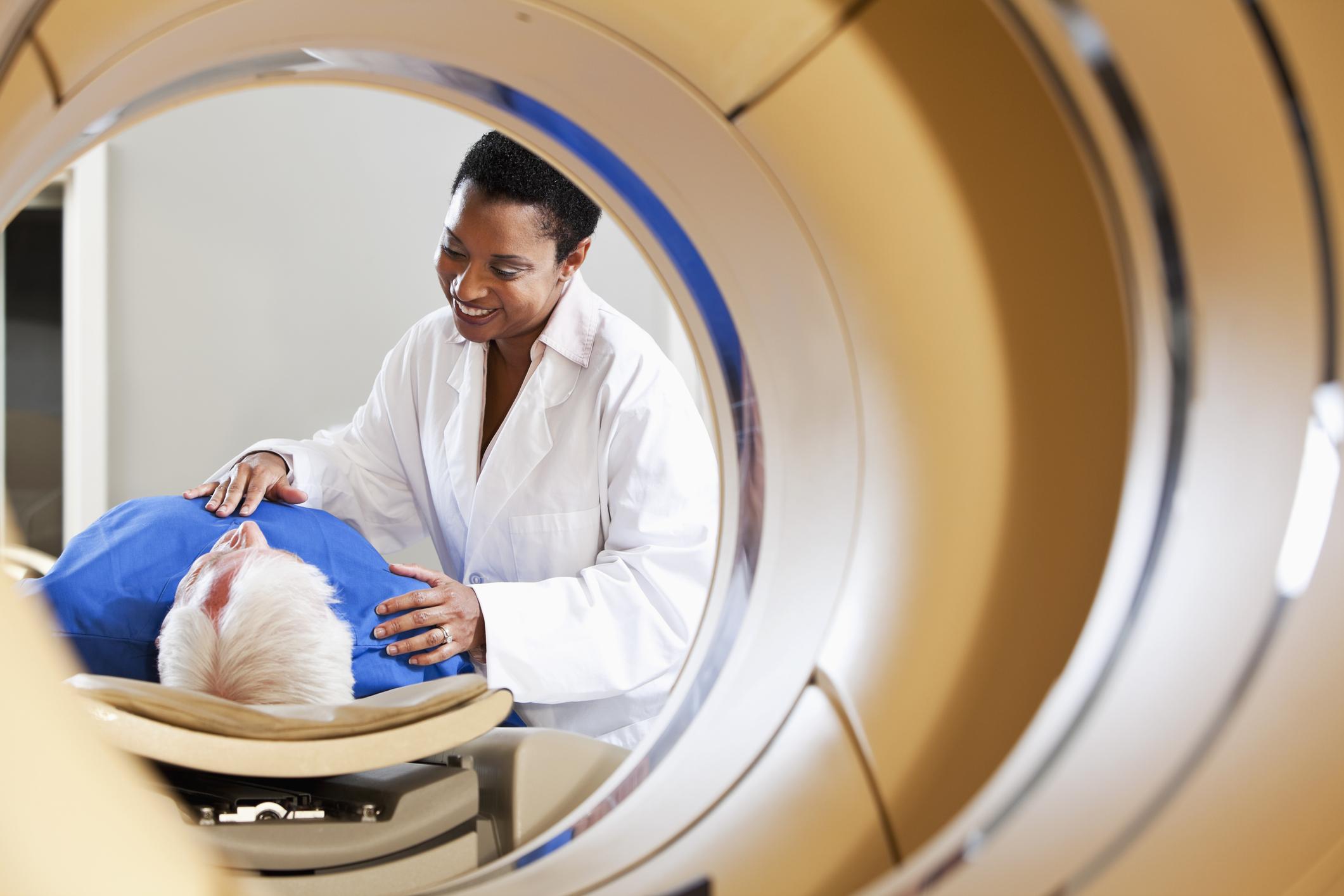
Using MRI to improve diagnosis
Following this work, in collaboration with Adam Boxer from UCSF, we published a paper in the journal JAMA Network Open to investigate the diagnostic value of MRI to identify patients with Progressive Supranuclear Palsy (PSP) and Corticobasal Degeneration (CBD)—two under-recognized causes of dementia. MRI is an imaging biomarker that is widely available for the diagnosis of neurodegenerative diseases. It has a lower cost and higher accessibility compared to other biomarkers. In this study, we applied different methods to quantify cerebral atrophy (loss of brain cells) on MRI at first diagnosis. We found that the combination of cortical and subcortical measures of atrophy had excellent accuracy in differentiating between diseases.
Sex differences in frontotemporal dementia
Despite the vast potential of MRI-based biomarkers to advance the diagnosis of neurodegenerative dementias, more work is needed to ensure their standardization and reproducibility. The effect of biological sex has emerged as a significant modifier of disease presentation and prognosis at the individual level, as we have shown in a recent paper published in Alzheimer's & Dementia: The journal of the Alzheimer's Association. The Alzheimer's Association-funded project titled "Unveiling the impact of biological sex along sporadic and genetic FTLD" will investigate this crucial topic and help us understand why women cope better with disease than men.
Overall, these data add to the increasing body of evidence supporting the value of MRI-based biomarkers as affordable and scalable tools to improve the recognition and diagnosis of neurodegenerative dementias worldwide, paving the way for the development of future disease-modifying therapies.
References:
Illán-Gala I, Montal V, Borrego-Écija S, et al. Cortical microstructure in primary progressive aphasia: a multicenter study. Alzheimers Res Ther. 2022;14(1):27. doi:10.1186/s13195-022-00974-0
Illán-Gala I, Nigro S, VandeVrede L, et al. Diagnostic Accuracy of Magnetic Resonance Imaging Measures of Brain Atrophy Across the Spectrum of Progressive Supranuclear Palsy and Corticobasal Degeneration. JAMA Netw Open. 2022;5(4):e229588. doi:10.1001/jamanetworkopen.2022.9588
Illán-Gala I, Falgàs N, Friedberg A, et al. Diagnostic Utility of Measuring Cerebral Atrophy in the Behavioral Variant of Frontotemporal Dementia and Association With Clinical Deterioration. JAMA Netw Open. 2021;4(3):e211290. doi:10.1001/jamanetworkopen.2021.1290
Illán‐Gala I, Casaletto KB, Borrego‐Écija S, et al. Sex differences in the behavioral variant of frontotemporal dementia: A new window to executive and behavioral reserve. Alzheimers Dement. 2021;17(8):1329-1341. doi:10.1002/alz.12299
Authors

Ignacio Illán-Gala, MD, PhD
Neurologist
GBHI Members Mentioned

Neus Falgàs, MD, PhD
Neurologist

Marilu Gorno Tempini, MD, PhD
Professor of Neurology and Psychiatry

Howie Rosen, MD
Professor of Neurology

Lea Tenenholz Grinberg, MD, PhD
Neuropathologist and Neuroscientist

Salvo Spina, MD, PhD
Associate Professor of Neurology

Bruce Miller, MD
Founding Director, University of California, San Francisco

Bill Seeley, MD
Professor of Neurology and Pathology